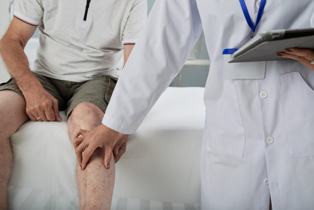
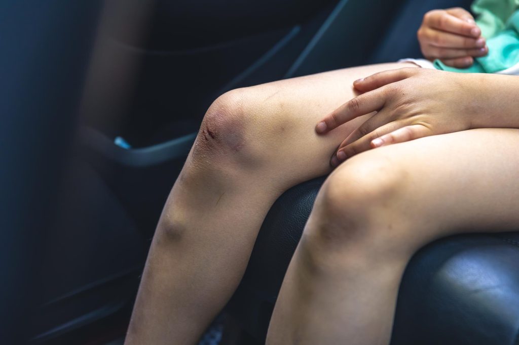
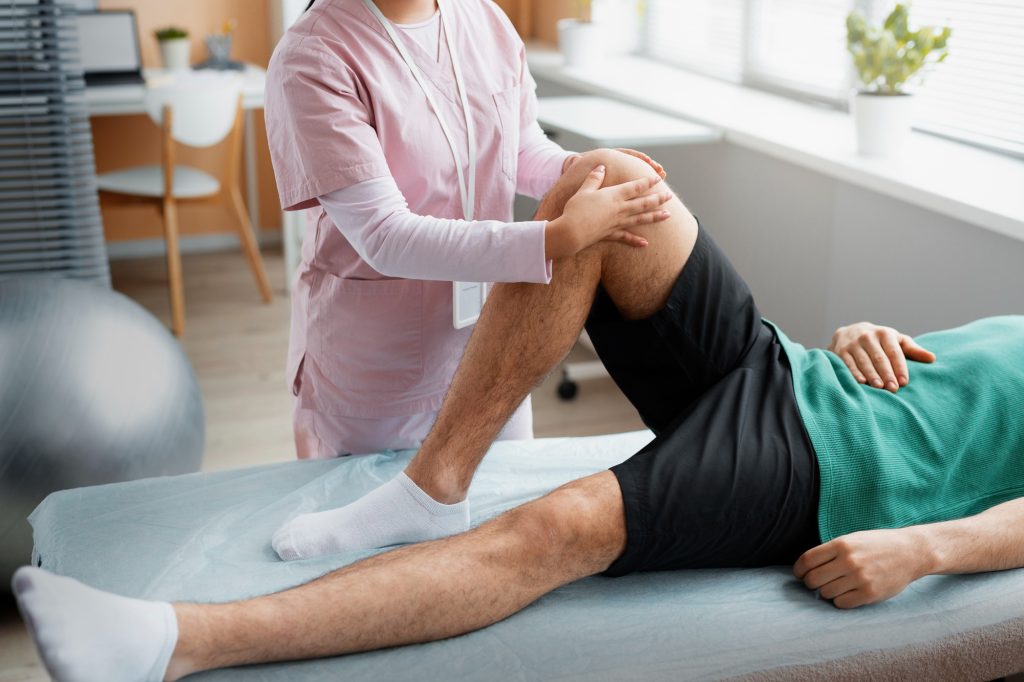
A swollen, painful knee can make walking, climbing stairs, and even standing feel impossible. When excess fluid accumulates in the knee joint, a condition called joint effusion, it can indicate arthritis, injury, infection, or other underlying issues. Knee arthrocentesis, commonly known as joint aspiration, is a minimally invasive procedure that removes this fluid, providing rapid relief and valuable diagnostic information.
At Joint Health Solutions in Charlotte, we perform knee arthrocentesis using ultrasound guidance to ensure precision, safety, and optimal outcomes. Our team—led by Dr. Jeffrey Galvin and Ariel Curtis, FNP-C—uses this procedure both therapeutically, to relieve pressure and pain, and diagnostically, to analyze fluid for infection, crystals, or inflammation. When appropriate, we may combine aspiration with a therapeutic injection such as corticosteroid to further reduce inflammation.
Medically reviewed by: Dr. Jeffrey Galvin & Ariel Curtis, FNP-C
Knee arthrocentesis is a procedure in which a sterile needle is inserted into the knee joint space to remove excess synovial fluid. The fluid may be:
The procedure serves two main purposes:
When needed, we can follow aspiration with an injection of corticosteroid to calm inflammation or obtain a sample for laboratory analysis.
The knee joint is lined by a synovial membrane that produces synovial fluid, a clear viscous fluid that lubricates and nourishes the joint. Under certain conditions, the membrane produces excessive fluid, leading to effusion. Common causes include:
| Cause | Description |
|---|---|
| Osteoarthritis | Inflammatory flare-up; fluid is typically clear to straw-colored. |
| Rheumatoid arthritis | Autoimmune inflammation; fluid may be cloudy. |
| Gout or pseudogout | Crystals cause acute inflammation; fluid is often turbid and contains crystals. |
| Septic arthritis | Bacterial infection; fluid may be purulent with a high white blood cell count and requires urgent treatment. |
| Trauma | Fracture, ligament tear, or meniscus injury; fluid may be bloody. |
| Bursitis | Fluid may collect outside the joint such as the prepatellar or pes anserine bursa. |
Arthrocentesis helps differentiate these conditions and guides treatment.
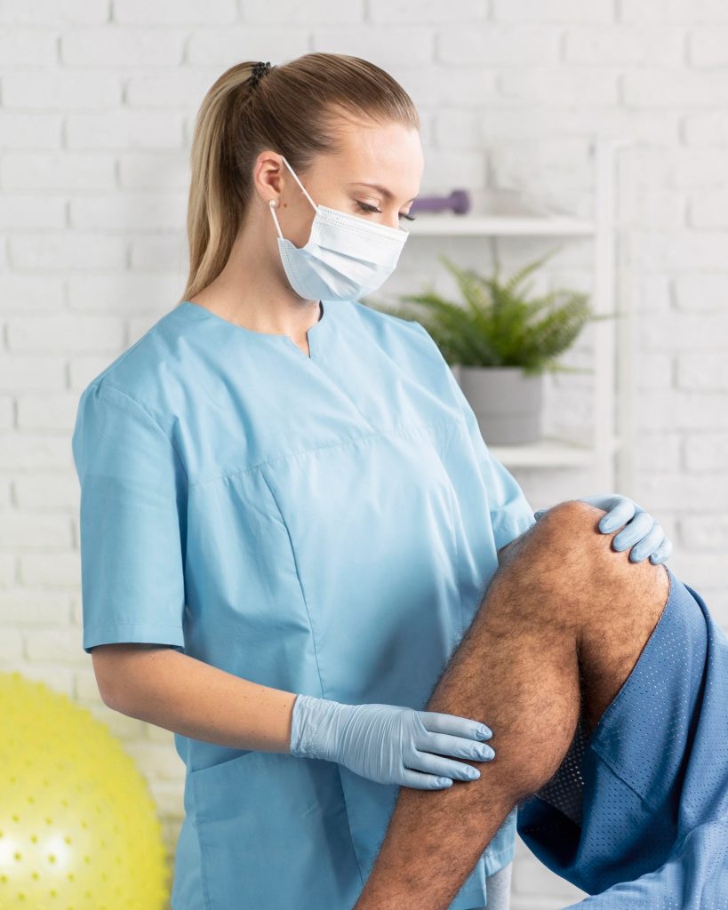
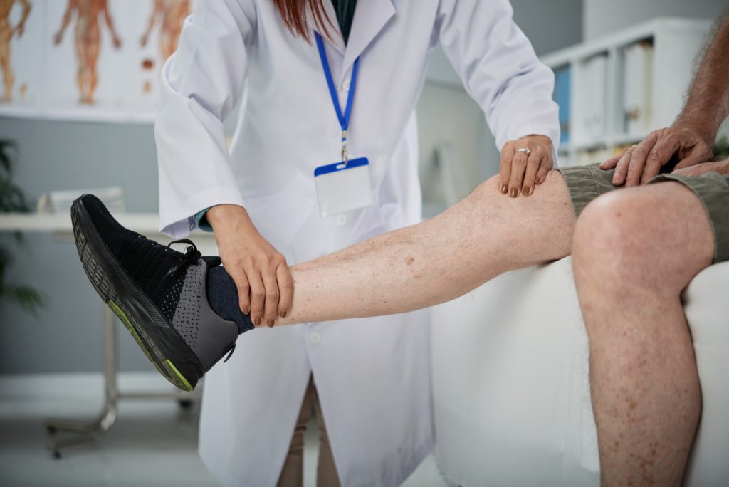
Your provider reviews your medical history, performs a physical exam, and assesses the knee for swelling, warmth, redness, and range of motion. We may also review prior imaging such as X-ray or MRI.
We use musculoskeletal ultrasound to confirm the presence of effusion, identify the best entry point, avoid blood vessels and nearby structures, and ensure the needle is placed accurately within the joint space.
The skin over the aspiration site is cleansed with an antiseptic and a sterile drape is placed. If desired, a local anesthetic may be used to numb the area.
A sterile needle is inserted into the joint space under ultrasound visualization and fluid is withdrawn into a syringe. The amount can range from a few milliliters to more than 100 mL in cases of large effusions.
If appropriate, a corticosteroid may be injected into the joint after aspiration to reduce inflammation and help prevent rapid re-accumulation of fluid.
The needle is removed and a small bandage is applied. You may be advised to rest the knee for 24–48 hours and apply ice. If fluid is sent for analysis, results typically return in 1–3 days for preliminary testing.
When fluid is sent to a laboratory, it may be evaluated for:
| Test | What It Detects |
|---|---|
| Appearance | Color and clarity such as clear, cloudy, purulent, or bloody. |
| Cell count & differential | White blood cell count helps differentiate inflammatory from septic arthritis. |
| Crystal analysis | Urate crystals in gout or calcium pyrophosphate crystals in pseudogout. |
| Gram stain & culture | Bacteria and antibiotic sensitivities when infection is suspected. |
| Glucose, protein, LDH | Additional markers of inflammation and joint pathology. |
These results help guide treatment, whether that means antibiotics for infection, urate-lowering therapy for gout, or anti-inflammatory management for osteoarthritis.
Knee arthrocentesis is very safe when performed by experienced providers using sterile technique and ultrasound guidance. Potential risks include:
We discuss all risks and benefits before the procedure.
| Procedure | Purpose |
|---|---|
| Arthrocentesis (aspiration) | Removes fluid, relieves pressure, and obtains a sample for diagnosis. |
| Corticosteroid injection | Reduces inflammation after aspiration or as a standalone treatment. |
| Hyaluronic acid injection | Lubricates the joint for osteoarthritis; usually performed when effusion is minimal. |
| PRP injection | Regenerative therapy; may be delayed until after effusion is controlled. |
At Joint Health Solutions, we often combine arthrocentesis with a corticosteroid injection to provide both immediate and sustained relief.
Expert Providers
Dr. Jeffrey Galvin and Ariel Curtis, FNP-C, have extensive experience in ultrasound-guided joint procedures.
Ultrasound Guidance
Ensures accurate needle placement, improves safety, and maximizes fluid yield.
Sterile Technique
Strict infection control protocols are followed.
Comprehensive Care
We combine aspiration with diagnostic analysis, therapeutic injections, and a long-term management plan.
Convenient Charlotte Location
Serving patients from Charlotte, Huntersville, Concord, Matthews, and surrounding areas.
Most patients feel a brief pinch from the local anesthetic and then pressure as the needle enters the joint. The procedure is generally well tolerated. After the aspiration, mild soreness may occur for a day or two.
The amount varies. Some patients have only a few milliliters, while others may have more than 100 mL of fluid. Removing a large effusion can provide dramatic relief.
The aspiration itself takes only a few minutes. Including preparation, ultrasound evaluation, and post-procedure care, the entire appointment typically lasts 20–30 minutes.
It depends on the underlying cause. If the cause is treated, such as infection, gout flare, or an osteoarthritis flare, fluid may not re-accumulate. If the underlying condition persists, fluid may return. In some cases, we combine aspiration with a corticosteroid injection to prolong relief.
Not always. If the diagnosis is already clear, such as known osteoarthritis with a non-inflammatory effusion, we may aspirate for therapeutic purposes only. If infection, gout, or inflammatory arthritis is suspected, we send fluid for analysis.
It depends on the type of blood thinner and the risk of bleeding. Some patients may need to temporarily hold their medication with approval from their prescribing physician, while others may proceed with caution. We evaluate each case individually.
Yes. After aspiration, we often inject a corticosteroid to reduce inflammation and prevent rapid re-accumulation. In some cases, we may aspirate first and schedule PRP therapy later, once acute inflammation is controlled.
Signs that aspiration may be beneficial include significant swelling that limits bending or straightening, a painful tense feeling in the knee, redness and warmth, or failure to improve with rest, ice, and oral anti-inflammatories.
Ultrasound guidance improves accuracy, especially in patients with complex anatomy or small effusions. It also helps avoid blood vessels and ensures the needle enters the correct compartment. At Joint Health Solutions, we use ultrasound for all joint aspirations.
If fluid is sent to a lab, preliminary results such as cell count and crystal analysis are often available within 24–48 hours. Culture results may take 3–5 days. We will contact you with results and discuss any needed treatment changes.
If your knee is swollen, painful, and limiting your mobility, schedule a consultation for knee arthrocentesis at Joint Health Solutions in Charlotte. We will help relieve your symptoms and determine the underlying cause.