
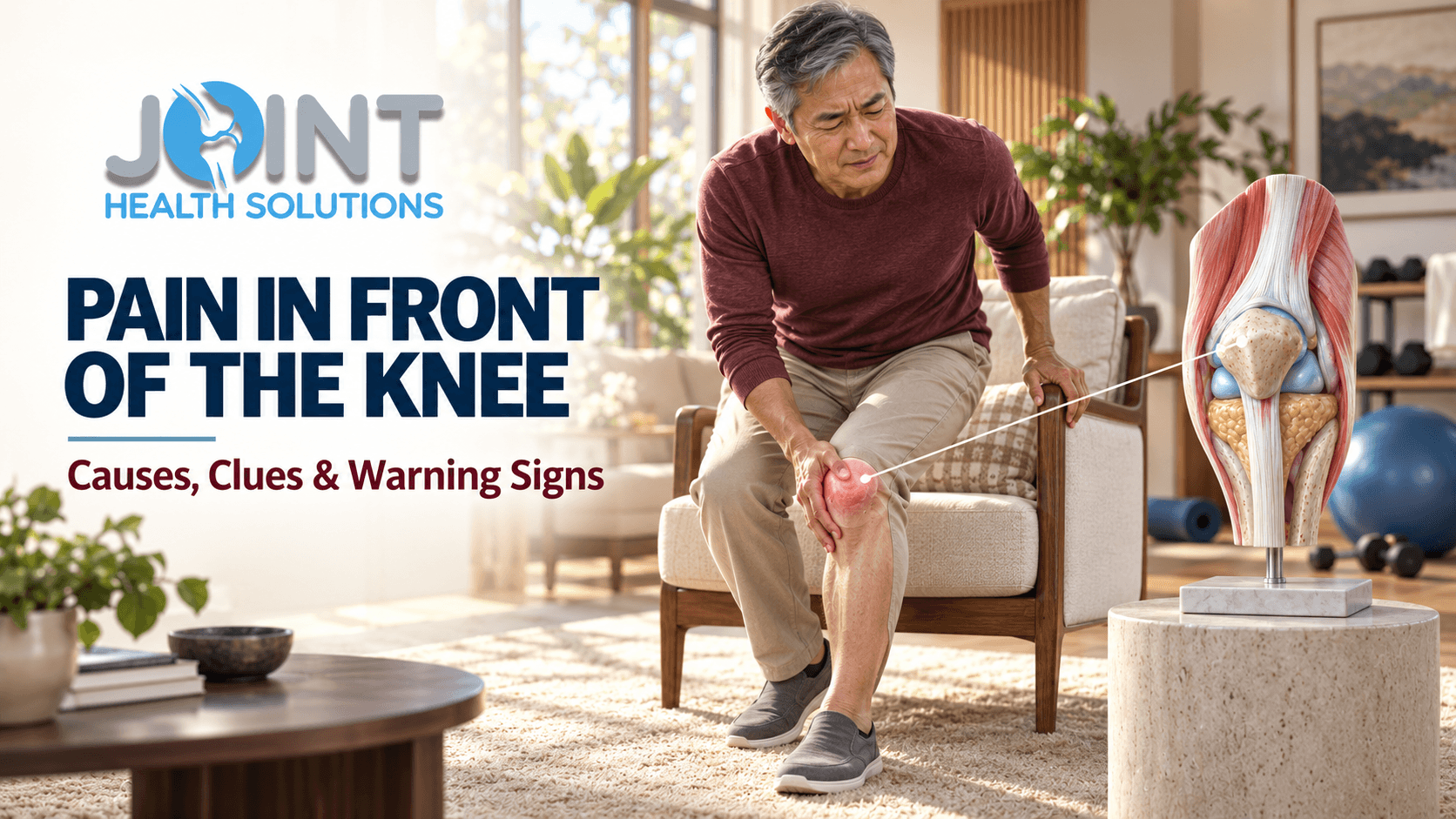
Pain in Front of the Knee: Causes, Location Clues, Warning Signs, and Evaluation
Pain in front of the knee—also called anterior knee pain—can come from the kneecap joint, patellar or quadriceps tendon, a bursa, the fat pad beneath the kneecap, arthritis, or an injury. The exact spot and the movements that provoke pain provide useful clues, but they cannot confirm the diagnosis by themselves.
This guide explains what may cause pain around, above, or below the kneecap; how stairs, squats, sitting, running, jumping, and kneeling change the possibilities; which symptoms need faster care; and how evaluation and treatment should be matched to the actual problem.
Quick Answer: What Causes Pain in Front of the Knee?
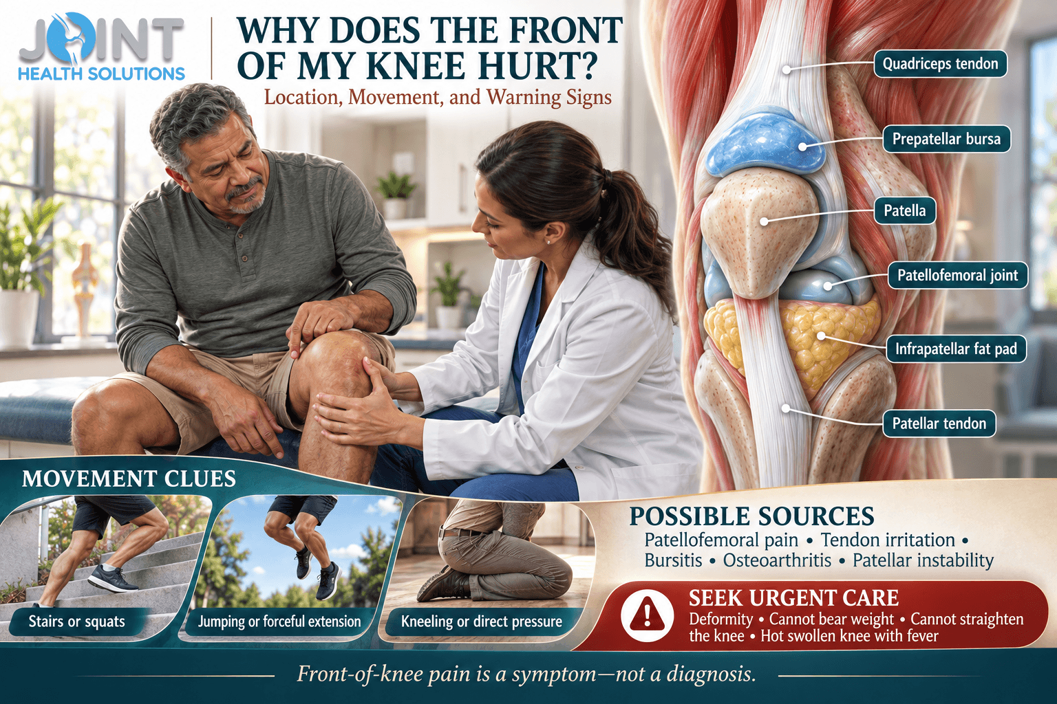
The most likely causes depend on the pain location and pattern. Pain around or behind the kneecap during stairs, squats, running, or prolonged sitting may fit patellofemoral pain. Focal pain just below the kneecap that increases with jumping can involve the patellar tendon. Pain above the kneecap may involve the quadriceps tendon. Direct tenderness and swelling over the kneecap can occur with prepatellar bursitis, while gradual pain and stiffness in an older adult may involve patellofemoral osteoarthritis.
Seek urgent care after a significant injury if the knee is deformed, you cannot bear weight, you cannot actively straighten it, or swelling develops rapidly. A hot, markedly swollen knee with fever or feeling ill also needs urgent assessment. New calf swelling plus chest pain, trouble breathing, fainting, or coughing blood is an emergency—call 911.
What Structures Are at the Front of the Knee?
The patella, or kneecap, sits within the quadriceps tendon and glides through a groove at the end of the femur as the knee bends and straightens. Its undersurface and the femoral groove are covered with articular cartilage. The joint between these surfaces is called the patellofemoral joint.
Above the kneecap, the quadriceps tendon connects the thigh muscles to the patella. Below it, the patellar tendon connects the patella to the tibia. Small fluid-filled bursae reduce friction around the front of the knee. The infrapatellar fat pad lies beneath and behind the patellar tendon and contains pain-sensitive tissue. The synovial lining, supporting retinacula, nearby bone, and skin can also produce symptoms.
Because these structures sit close together, “kneecap pain” is not one diagnosis. Pain centered behind the patella during a squat differs from pinpoint tenderness at the tendon, visible swelling directly over the kneecap, or sudden loss of the ability to straighten the leg after a fall. A careful location-and-load history helps separate those patterns.
Front-of-Knee Pain Location Clues
| Where it hurts | Possible source | Helpful accompanying clues |
|---|---|---|
| Around or behind the kneecap | Patellofemoral pain or patellofemoral osteoarthritis | Stairs, squats, running, or sitting with the knee bent may provoke pain |
| Just below the kneecap | Patellar tendon or infrapatellar fat pad | Jumping and forceful knee extension may load the tendon; full extension may irritate the fat pad |
| Above the kneecap | Quadriceps tendon | Pain with resisted straightening, stairs, squats, or a recent load increase |
| Directly over the kneecap | Prepatellar bursa, skin, or patella | Kneeling, direct impact, visible swelling, warmth, or focal bone tenderness |
| Inner edge of the kneecap | Patellofemoral tissues, plica, or prior patellar instability | Clicking, a catching sensation, or a history of the kneecap shifting |
| Upper shin bump in an adolescent | Tibial tubercle traction irritation | Sports-related pain and tenderness during a growth period |
Common Causes of Pain in Front of the Knee
Patellofemoral pain
Patellofemoral pain describes discomfort in front of or around the kneecap. It often develops gradually and is associated with activities that repeatedly load a bent knee, including stairs, squatting, running, jumping, and prolonged sitting. A recent increase in training frequency, distance, intensity, hills, or load may be relevant. It affects athletes and nonathletes.
The cause is rarely one simple flaw. Training load, tissue sensitivity, hip and knee strength, movement control, recovery, footwear, and individual anatomy can interact. Clicking or grinding may occur, but sound does not prove cartilage damage. Conversely, a quiet knee can still be painful. The dedicated guide to knee clicking and popping explains when joint noise is more concerning.
Patellar tendinopathy
The patellar tendon transmits force from the quadriceps through the kneecap to the shin. Tendinopathy commonly causes focal pain at the lower tip of the patella and is often linked with jumping, sprinting, rapid deceleration, or repeated heavy knee loading. Symptoms may warm up during activity and return afterward, but this pattern varies.
Tendon pain is not simply inflammation, and complete rest does not automatically restore load capacity. Management often requires a temporary reduction in provocative load followed by structured, progressive strengthening. Sudden pain, a palpable gap, major bruising, or inability to actively straighten the knee raises concern for a partial or complete tendon tear rather than routine tendinopathy.
Quadriceps tendon pain
The quadriceps tendon attaches above the kneecap. Irritation may produce localized pain during squats, stairs, rising from a low chair, running, or resisted knee extension. A rapid increase in training or a return to high-load activity after time away may contribute. In adults with sudden trauma, especially a forceful contraction while the knee is bent, an extensor-tendon tear must be considered if straightening the leg becomes difficult or impossible.
Prepatellar bursitis
The prepatellar bursa is a small sac between the skin and kneecap. Repeated kneeling or a direct blow can irritate it, causing a distinct pocket of swelling over the front of the patella. Some people can still move the joint reasonably well even though kneeling is painful.
A bursa can also become infected. Increasing warmth, redness or skin-color change, drainage, fever, chills, or feeling unwell requires prompt medical assessment. Do not attempt to drain a swollen bursa at home. The clinician may need to distinguish bursitis from joint infection, cellulitis, gout, or another cause.
Infrapatellar fat-pad irritation
The infrapatellar fat pad is a richly innervated structure under and behind the patellar tendon. It can become sensitive after a direct impact, surgery, repeated knee extension, or a period of altered movement. Pain is usually at the lower front of the knee and may be worse when the knee is forcefully straightened or held in extension. It can resemble patellar tendon or patellofemoral pain, so self-diagnosis based on tenderness alone is unreliable.
Patellofemoral osteoarthritis
Osteoarthritis can affect the compartment behind the kneecap, the inner or outer tibiofemoral compartment, or several areas at once. Patellofemoral osteoarthritis may cause front-of-knee pain, stiffness, swelling, crepitus, and difficulty with stairs, slopes, squatting, or standing from a low seat. Symptoms and imaging do not always match, so an X-ray finding should be interpreted alongside function and examination.
This article does not replace the broader knee osteoarthritis guide or treatment-specific pages. Its purpose is to explain anterior pain location. Osteoarthritis care may include education, exercise, weight-management support when relevant, medication review, and selected procedures, but no option can promise permanent relief or cartilage regrowth.
Patellar instability or dislocation
The kneecap can shift partly or completely out of its groove, most often toward the outer side. A first episode may follow a pivot, awkward landing, or direct force and can produce sudden pain, swelling, a visible deformity, or a feeling that the kneecap moved. Even if it returns to position on its own, a first-time traumatic episode deserves assessment for associated bone, cartilage, ligament, and alignment factors.
Recurrent apprehension or instability is different from general knee weakness. Do not repeatedly push the kneecap sideways to test it. If the knee gives way without an obvious kneecap shift, see the separate article on knee buckling and giving way.
Plica, cartilage, bone, and referred pain
A fold in the joint lining called a plica may become irritated and cause pain or a snapping sensation near the inner front of the knee. A focal cartilage or bone injury, patellar fracture, stress injury, inflammatory arthritis, gout, or infection can also cause anterior symptoms. In adolescents, traction irritation near the tibial tubercle or lower patella may be related to growth and sports loading.
Not every sensation at the front of the knee begins there. Hip weakness or pain, lumbar nerve irritation, altered ankle mechanics, or gait changes may influence knee loading or refer symptoms. Numbness, tingling, back pain, hip pain, or pain that does not behave like a local knee problem broadens the examination.
What Different Activity Patterns May Suggest
| When pain occurs | Patterns to consider | Details worth reporting |
|---|---|---|
| Stairs, squats, or low chairs | Patellofemoral pain, osteoarthritis, or extensor-tendon loading | Exact location, depth of bend, weakness, swelling, and whether ascent or descent is worse |
| Sitting with the knee bent | Patellofemoral joint irritation | Time to onset, relief with straightening, stiffness, and clicking |
| Jumping, sprinting, or quick deceleration | Patellar or quadriceps tendon, patellofemoral tissues | Focal tendon tenderness, training change, sudden pop, and ability to extend the knee |
| Kneeling | Prepatellar bursa, skin, or patella | Visible swelling, warmth, recent cut, repeated pressure, or direct impact |
| After a pivot, fall, or collision | Patellar instability, tendon tear, fracture, cartilage injury, or other internal injury | Deformity, rapid swelling, pop, inability to bear weight, or loss of active extension |
When Is Pain in Front of the Knee Urgent?
Most gradual anterior knee pain is not an emergency. Urgency changes when trauma, loss of function, infection, circulation, or a possible blood clot is involved. Do not keep exercising to test a knee that is deformed, unstable, rapidly swelling, or unable to support you.
Seek emergency or urgent assessment for:
- A visibly deformed knee or kneecap after injury
- Inability to bear weight after significant trauma
- Inability to actively straighten the knee or keep the straight leg lifted
- Rapid, substantial swelling after a pop, pivot, fall, or collision
- A foot that becomes cold, pale, blue, numb, or weak
- A hot, markedly swollen knee with fever, chills, drainage, or feeling acutely ill
- New one-sided calf or leg swelling with chest pain, shortness of breath, fainting, coughing blood, or a rapid heartbeat—call 911
Arrange prompt evaluation for recurrent kneecap shifting, repeated giving way, persistent swelling, a knee that becomes mechanically stuck, progressive weakness, focal bone pain, or symptoms that disturb sleep or limit walking and daily activity. Pain at night has its own differential; see the guide to knee pain at night for that separate intent.
What Can You Do Before an Evaluation?
For mild, nontraumatic pain without warning signs, reduce the movement or training dose that sharply increases symptoms. That may mean temporarily shortening runs, limiting deep loaded squats, reducing jumping volume, using a higher chair, or taking breaks from prolonged knee flexion. Comfortable walking or low-impact motion may be reasonable if it does not increase pain, swelling, limping, or instability.
A wrapped cold pack can help short-term soreness or swelling; protect the skin and use brief sessions. Compression and elevation may be reasonable for mild swelling if they do not cause numbness, color change, or more pain. Medication is not safe for everyone. Kidney disease, ulcers, bleeding risk, cardiovascular disease, pregnancy, allergies, and interactions can change whether an anti-inflammatory or other pain reliever is appropriate.
Avoid forcefully pressing, grinding, or shifting the kneecap to reproduce symptoms. Do not repeatedly hop, squat, or extend the knee through sharp pain as a home “test.” A generic brace, taping method, or online exercise may aggravate the wrong condition. Seek evaluation when function worsens or a reasonable period of load modification does not produce improvement.
How Clinicians Evaluate Anterior Knee Pain
The history should establish when symptoms began, the exact location, whether onset was sudden or gradual, the movement that provokes pain, recent changes in activity, and how the knee behaves afterward. Swelling timing, a pop, instability, locking, fever, skin injury, previous dislocation, prior surgery, medication use, and hip or back symptoms can change the differential.
During the examination, a clinician may compare both legs for swelling, warmth, alignment, gait, motion, tenderness, patellar movement, tendon continuity, strength, and the ability to actively extend the knee. Squatting, stepping, or another functional task may be observed when safe. Hip strength and motion, ankle mobility, neurologic findings, and circulation may also be assessed.
When imaging or testing may help
Imaging should answer a clinical question, not replace the examination. X-rays can assess fracture, alignment, patellar position, bone changes, and osteoarthritis. Ultrasound can evaluate selected superficial tendons, bursae, and fluid collections when performed by an experienced clinician. MRI may be useful when a significant tendon, cartilage, bone, ligament, or other internal injury is suspected and the result would change management.
Not every case of patellofemoral pain needs advanced imaging. Conversely, a normal X-ray does not exclude every cartilage, tendon, or early bone stress problem. Blood tests or joint-fluid analysis may be needed when infection, crystal arthritis, or systemic inflammation is suspected. The article on a first non-surgical joint evaluation explains how to prepare and what information to bring.
How Treatment Changes With the Diagnosis
“Front knee pain” is a location, not a treatment plan. A gradual overload condition, inflamed bursa, arthritic joint, unstable kneecap, tendon tear, fracture, and infection require different care. The safest plan connects the diagnosis, symptom severity, daily demands, medical history, and personal goals.
| Clinical pattern | Possible care components | Important limitation |
|---|---|---|
| Patellofemoral pain | Education, load adjustment, progressive hip and knee exercise, movement and activity planning | No single alignment finding, brace, or exercise explains or fixes every case |
| Patellar or quadriceps tendinopathy | Temporary load reduction followed by progressive tendon loading and gradual return to sport or work | Complete tears and routine tendinopathy are not managed the same way |
| Prepatellar bursitis | Reduce pressure, protect the area, evaluate persistent fluid; antibiotics or drainage when infection is diagnosed | A hot or draining bursa should not be treated as simple overuse at home |
| Patellofemoral osteoarthritis | Exercise, education, weight-management support when relevant, medication review, selected bracing or procedure | Benefits vary; no nonsurgical treatment guarantees permanent relief or cartilage regrowth |
| Instability, fracture, or tendon tear | Protection, appropriate imaging, specialist input, and rehabilitation or surgery when indicated | Continuing activity or receiving a pain procedure cannot restore disrupted structure |
Physical therapy and progressive loading
Physical therapy may address quadriceps and hip strength, mobility, movement tolerance, balance, gait, work tasks, and a graded return to activity. Exercise dosage matters. A program that is appropriate for gradual patellofemoral pain may be unsafe immediately after a fracture, dislocation, or suspected tendon tear.
Braces, taping, medication, and injections
Taping or a brace may provide short-term support for selected patellofemoral or instability patterns, but fit and purpose matter. Medication may be considered after contraindications and interactions are reviewed. An injection may be discussed for selected arthritis or inflammatory diagnoses, but it is not automatically appropriate because pain is at the front of the knee.
An injection cannot repair a ruptured tendon, reduce an unreduced dislocation, stabilize a major structural injury, heal a fracture, or treat an infection. Expected benefit, alternatives, evidence, and risks should be discussed for the specific diagnosis. Insurance coverage varies by plan, medical necessity, authorization requirements, and service; it must be verified individually.
What Should You Track Before an Appointment?
- Point with one finger to the most painful spot: above, below, over, around, or behind the kneecap
- Record which movements provoke pain and how long symptoms last afterward
- Note recent changes in exercise, work, hills, stairs, footwear, or kneeling
- Describe any fall, direct blow, pivot, pop, or kneecap-shifting episode
- Track swelling timing, warmth, redness or skin-color change, locking, or instability
- Note whether you can actively straighten the knee and walk without limping
- Bring relevant imaging, reports, medications, allergies, and insurance information
A short symptom record is useful; repeatedly provoking sharp pain is not. If swelling changes over time, a photo may help document it. Do not recreate a jump, deep squat, kneecap shift, or unstable movement for a video.
Evaluation for Pain in Front of the Knee in Charlotte, NC
Joint Health Solutions evaluates knee pain, swelling, stiffness, instability, injury-related symptoms, and activity limitations at 9350 Benfield Rd, #109, Charlotte, NC 28269. The first goal is to determine the most plausible pain source and whether education, activity modification, physical therapy, imaging, bracing, a procedure, or referral is appropriate.
An appointment does not guarantee that imaging, an injection, or a brace will be recommended. Symptoms suggesting a fracture, tendon rupture, unreduced dislocation, infection, blood clot, or circulation problem may require urgent hospital-based care or specialty referral beyond an outpatient joint-pain visit. Call 704-833-3566 for routine scheduling questions; call 911 for a medical emergency.
Frequently Asked Questions About Pain in Front of the Knee
What is the most common cause of pain in front of the knee?
Patellofemoral pain is a common cause, especially when discomfort occurs around or behind the kneecap during stairs, squats, running, or prolonged sitting. It is not the only cause. Age, trauma, exact location, swelling, tenderness, instability, and the activities that reproduce symptoms help distinguish tendon, bursa, arthritis, fat-pad, bone, and kneecap-instability problems.
Why does the front of my knee hurt when I squat?
Squatting increases demand on the patellofemoral joint and knee-extensor system. Patellofemoral pain, patellofemoral osteoarthritis, or patellar and quadriceps tendon pain may become noticeable. Depth, load, volume, technique, recent training changes, and tissue capacity all matter. Sharp pain, swelling, instability, or persistent loss of function warrants evaluation.
Why does my kneecap hurt going down stairs?
Descending stairs requires controlled quadriceps work while the knee is bent, which increases patellofemoral demand. Patellofemoral pain and patellofemoral osteoarthritis are possibilities, but tendon pain or another condition may overlap. The symptom is not a diagnosis, and the precise location, swelling, age, injury history, and examination remain important.
Can sitting too long cause pain around the kneecap?
Prolonged sitting with the knee bent can provoke patellofemoral symptoms in some people. Straightening the leg or changing position may reduce discomfort. Sitting does not prove cartilage damage, and persistent pain should not automatically be blamed on posture. New swelling, locking, instability, or significant motion loss changes the evaluation.
How can I tell patellar tendon pain from patellofemoral pain?
Patellar tendon pain is often focal at the lower tip of the kneecap and closely linked with jumping or forceful knee extension. Patellofemoral pain is usually more diffuse around or behind the kneecap and may be provoked by stairs, squats, running, or sitting. The patterns can overlap, and pressing on the area alone is not a reliable diagnosis.
Does grinding in the front of the knee mean arthritis?
No. Crepitus can occur with or without pain and does not by itself diagnose arthritis or cartilage injury. Arthritis becomes more plausible when the overall pattern includes age-related risk, stiffness, swelling, reduced motion, activity-related pain, and compatible imaging. Treatment decisions should not be based on sound alone.
Should I exercise with anterior knee pain?
Appropriate movement and progressive strengthening are often part of care for gradual, nontraumatic patellofemoral or tendon pain. The exercise type and dose should not sharply increase symptoms or cause worsening swelling, limping, or loss of function. Stop and seek assessment after trauma, a pop with weakness, inability to straighten the knee, deformity, or other warning signs.
Do I need an MRI for pain in front of the knee?
Not routinely. Many cases can be assessed through history, examination, and—when indicated—X-rays. MRI may help when a major tendon, cartilage, bone, ligament, or other internal injury is suspected, symptoms persist despite appropriate care, or the result would change management. Imaging should answer a specific question.
When should swelling over the kneecap be checked?
Prompt assessment is appropriate when swelling follows significant trauma, grows quickly, limits function, or occurs with warmth, redness or skin-color change, drainage, fever, chills, or feeling ill. Those features can indicate fracture, bleeding, infection, or another condition rather than uncomplicated pressure-related bursitis.
Can an injection treat pain in front of the knee?
Only selected diagnoses may justify an injection, and expected benefits and risks vary. An injection is not a default treatment for patellofemoral pain or tendon overload and cannot repair a torn tendon, correct instability, heal a fracture, or treat infection. A diagnosis-first evaluation should determine whether a procedure belongs in the plan.
Key Takeaway
Pain in front of the knee is best understood by combining exact location, provoking load, symptom timing, swelling, mechanical symptoms, trauma, and function. Patellofemoral pain is common, but tendon, bursa, arthritis, fat-pad, instability, bone, and infection-related causes need different care. Red flags and loss of function determine urgency; the diagnosis determines treatment.
Sources
- American Academy of Orthopaedic Surgeons: Patellofemoral Pain Syndrome
- American Academy of Orthopaedic Surgeons: Arthritis of the Knee
- American Academy of Orthopaedic Surgeons: Patellar Tendon Tear
- American Academy of Orthopaedic Surgeons: Prepatellar Bursitis
- American College of Radiology: Appropriateness Criteria for Chronic Knee Pain
- NHS: Septic Arthritis