

When dealing with chronic musculoskeletal pain, targeted joint and spinal injections can provide profound relief and help you reclaim your daily life. However, the success of any injection treatment relies heavily on one single factor: absolute anatomical precision. Delivering powerful therapeutic agents exactly where they need to go is essential—especially when targeting deep, anatomically complex, or structurally narrow joints like the hip, sacroiliac (SI) joint, or the delicate facet joints of the spine. Attempting to insert a medical needle into these deep spaces using only touch, surface landmarks, and clinical intuition—a method known as a “blind” or landmark-guided injection—presents clear limitations. Studies have shown that blind injections into deep anatomical structures can result in miss rates as high as 30% to 40%. When an injection misses its marks, patients continue to suffer from unmanaged pain, while valuable medications are wasted in surrounding tissues. To avoid these pitfalls, Joint Health Solutions utilizes fluoroscopy-guided injections. This live, image-guided approach provides real-time visualization of your joint anatomy, transforming a blind medical procedure into a pinpoint-accurate, highly visual treatment. Whether you are receiving advanced corticosteroids, hyaluronic acid visco-supplementation, or regenerative treatments like Platelet-Rich Plasma (PRP) therapy, our team utilizes fluoroscopy to ensure maximum safety, improved patient comfort, and optimal clinical outcomes.
At its core, fluoroscopy is an advanced form of medical imaging that functions like a continuous, real-time X-ray video on a high-definition monitor. While standard, conventional diagnostic X-rays capture a single, static image of your bones, fluoroscopy records dynamic movement. This allows an interventional specialist to see deep internal structures, track joint movement, and watch a medical needle advance through tissue layers toward its target.
The primary equipment utilized in these procedures is called a C-arm. Named for its unique structural “C” shape, this device features an X-ray generator on one end and an image intensifier detector on the other.
The C-arm rotates smoothly around an examination table, allowing the provider to view your joints from multiple angles and three-dimensional perspectives without requiring you to constantly shift your body during the procedure.
💡 Quick Anatomy of a C-Arm Fluoroscope
To understand how this real-time imaging system functions during your procedure, think of it as a vertical loop working in perfect synchronization:
The X-Ray Beam Source (Bottom): Positioned safely beneath the examination table, this component emits a precise, low-dose pulsed beam upward through the targeted joint.
The Patient on the Table (Center): You rest comfortably in a stabilized position while the pulsed radiation safely passes through the precise anatomical window being treated.
The X-Ray Intensifier Screen (Top): Positioned directly above you, this high-tech detector captures the upward beams and instantly converts them into a high-definition image.
The Continuous Live Video Feed (Output): The captured data is sent in real time to a medical monitor, allowing your specialist to guide the needle with millimeter-level accuracy.
During a typical live-guided procedure at our clinic, several events happen simultaneously:
The patient is positioned comfortably on a radiolucent (X-ray transparent) imaging table.
The C-arm is carefully aligned over the specific treatment area.
The provider tracks the monitor to guide the needle forward.
To confirm placement before injecting any medication, a tiny amount of contrast dye is introduced. If the dye flows smoothly throughout the joint capsule, it confirms the needle is perfectly positioned.
This dynamic process adds only a few minutes to the total appointment time, but it significantly improves accuracy and patient safety.
Entering a joint space is a highly detailed task. Many of the body’s deep joints are surrounded by networks of nerves, major blood vessels, tendons, and dense muscle tissue. If a needle is placed without image guidance, even a minor variance can lead to clinical issues:
According to clinical research published in the Pain Physician Journal, classic blind landmark-guided injections show error rates between 30% and 40% for deep spinal, pelvic, and hip structures. By utilizing fluoroscopic guidance, our providers reduce the margin of error to less than 5%. This high degree of accuracy ensures your treatment is delivered safely and effectively.
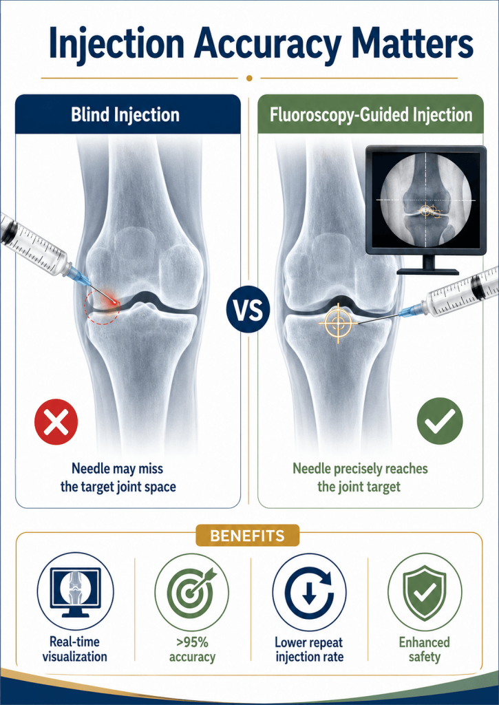
Fluoroscopy guidance is invaluable for treating deep joints that cannot be easily palpated or evaluated from the surface of the skin. The table below outlines the specific interventions that benefit most from this real-time imaging technology:
| Procedure Name | Typical Clinical Uses & Diagnoses | Why Fluoroscopy is Essential |
Hip Joint Injection | Advanced hip osteoarthritis, labral tears, and avascular necrosis. | The hip is a deep ball-and-socket joint beneath layers of muscle and fat; blind entry attempts miss the joint capsule roughly 30% of the time. |
Sacroiliac (SI) Joint Injection | Sacroiliac joint dysfunction, sacroiliitis, and localized lower back or buttock pain. | The SI joint is a narrow, irregular pathway; it is very difficult to access accurately without real-time visualization. |
Facet Joint Injection / Medial Branch Block | Spinal arthritis, facet joint syndrome, and chronic back pain. | These small joints link your vertebrae; fluoroscopy ensures the needle makes direct contact with the target bone structure while avoiding nearby spinal nerves. |
Epidural Steroid Injection (ESI) | Radicular pain, sciatica, lumbar disc herniations, and spinal stenosis. | ESIs require navigating the delicate epidural space; real-time guidance is critical to avoid the spinal cord and ensure safe medication delivery. |
Shoulder (Glenohumeral) Injection | Advanced shoulder arthritis, frozen shoulder (adhesive capsulitis), and severe stiffness. | Recommended when patient anatomy is altered or when prior blind injections have failed to provide relief. |
Knee Arthrocentesis (Fluid Aspiration) | Chronic joint effusion, diagnostic sampling, and post-traumatic swelling. | Useful when managing complex or severely scarred joint spaces where simple landmark assessment is insufficient. |
Note on Alternative Imaging Modalities: For more superficial structures, such as standard knee injections or subacromial shoulder bursitis treatments, we often utilize advanced musculoskeletal ultrasound guidance. This provides real-time visualization of soft tissues without any radiation exposure. Your provider will select the optimal imaging technology based on your unique anatomy and diagnosis.
To understand why image guidance has become a gold standard for interventional orthopedics, it helps to compare fluoroscopy directly against traditional, unguided landmark methods:
| Feature Matrix | Fluoroscopy-Guided Injections | Blind (Landmark) Injections |
| Average Accuracy Rate |
>95% precision across all deep structures | 60% to 80% (varies considerably by joint depth) |
| Real-Time Visualization |
Yes — Full video view of needle progression |
No — Performed based on touch and anatomical estimations |
| Contrast Spread Confirmation |
Yes — Confirms exact fluid distribution via dye |
No — Verification of fluid location is not possible |
| Repeat Injection Rate |
Extremely Low due to high initial success rates |
Higher due to missed targets or partial delivery |
| Patient Safety Profile |
Superior — Clear view avoids nerve pathways |
Moderate — Higher risk of clipping soft tissues |
| Total Procedure Time | Slightly longer (10–20 minutes overall) | Shorter (2–5 minutes total) |
| Radiation Exposure Profile | Minimal, strictly monitored low-dose exposure |
None |
While blind injections may save a couple of minutes upfront, that minor time savings is far outweighed by the accuracy, safety, and reliable pain relief provided by fluoroscopic visualization.
It is entirely natural to feel a bit anxious before undergoing any procedure involving needles or imaging equipment. Knowing exactly what happens before, during, and after your visit can help alleviate these concerns.
1. Before the Procedure
Your care begins well before you step into our procedure suite. During your initial consultation, your provider will thoroughly review your medical history, current medications, and any known allergies.
Medication Modifications: If you take prescription blood thinners or anti-platelet therapies, we will coordinate with your prescribing physician to help you temporarily stop them before your injection to minimize bruising or bleeding risks.
Dietary Guidelines: No fasting or special dietary restrictions are required unless you and your physician have decided to utilize conscious sedation (which is rare for routine joint procedures).
Wardrobe Tips: We recommend wearing loose, comfortable clothing to your appointment, though you may be asked to change into a clinical gown depending on the targeted joint.
2. During the Procedure
When you enter the treatment suite, our team focused on keeping you safe and comfortable:
Positioning: You will lie down on our specialized fluoroscopy table. Your positioning will depend on the target joint—for example, you’ll lie on your back for a hip injection, or on your stomach for a spinal facet joint procedure.
Sterile Preparation: The skin over the target joint is thoroughly cleansed with an advanced antiseptic solution to ensure a sterile environment.
Local Anesthesia: We apply a topical numbing spray or inject a small amount of local lidocaine to numb the skin, minimizing any discomfort from the therapy needle.
Target Identification: The C-arm is positioned, and initial quick images are taken to align the injection path with your anatomy.
Precision Advancement: Under live X-ray guidance, our specialist carefully guides the needle directly into the joint space.
Contrast Confirmation: When appropriate, a tiny droplet of contrast dye is introduced to confirm perfect placement on the monitor.
Medication Delivery: Once placement is confirmed, the therapeutic solution (corticosteroids, HA, or PRP) is smoothly delivered.
Completion: The needle is removed, and a small, clean bandage is applied over the site. The entire process typically takes just 10 to 20 minutes.
3. After the Procedure
Observation Window: You will rest comfortably in our recovery area for 5 to 10 minutes to ensure you feel well before leaving.
Transportation: Since conscious sedation is rarely used, most patients can safely drive themselves home after the visit.
Activity Levels: You can resume light, normal daily activities immediately. However, we advise avoiding strenuous workouts, heavy lifting, or high-impact exercise for the first 24 to 48 hours.
Managing Post-Injection Flares: If you received a corticosteroid injection, you may experience a temporary “cortisone flare”—a minor increase in localized aching that can last 24 to 48 hours. Applying ice packs and resting your joint will quickly resolve this discomfort. Always follow your provider’s specific recovery guidelines, such as avoiding over-the-counter NSAIDs after a cellular PRP treatment.
A frequent question patients ask is: “Is the radiation exposure from a fluoroscope safe?” While fluoroscopy does utilize ionizing radiation to generate its live video stream, the cumulative dose delivered during a standard joint injection is exceptionally low.
The typical radiation exposure ranges between 0.5 to 2 mSv per procedure. To provide some perspective on what those numbers mean in daily life, that dose is roughly equivalent to:
Our clinical team strictly adheres to the ALARA (As Low As Reasonably Achievable) safety framework. We protect our patients by employing specific, rigorous safety protocols:
⚠️ Pregnancy Warning: Ionizing radiation should generally be avoided during pregnancy to protect developing fetuses. If you are pregnant, suspect you might be, or are actively planning a pregnancy, please inform our clinical staff immediately before scheduling your appointment so we can discuss alternative options.
Image-guided treatments are valuable for managing conditions that have not responded well to standard physical therapies or oral medications:
Advanced Hip Osteoarthritis: Helps deliver anti-inflammatory relief deep into the hip joint capsule, which can also confirm whether the hip is the true source of your pain.
Sacroiliac Joint Dysfunction: Pinpoints the narrow SI joint spaces to relieve lower back and buttock pain that is often misdiagnosed as disc issues.
Spinal Facet Arthritis: Provides accurate delivery for medial branch blocks or direct facet joint treatments to relieve chronic spinal pain.
Severe Sciatica and Radiculopathy: Places epidural steroid therapies precisely around compressed nerve roots to calm nerve inflammation caused by herniated discs or spinal stenosis.
Overcoming Failed Blind Injections: If you have had landmark-guided injections in the past that failed to provide relief, it may have been due to a missed target. Fluoroscopy ensures the medication is placed correctly.
No, the fluoroscopy imaging portion of the treatment is completely painless—the C-arm moves around you without touching you. The injection itself feels very similar to a routine blood draw or vaccine, characterized by a brief pinch and pressure as the skin is numbed.
The exposure is very low, typically between 0.5 and 2 mSv per injection. This is comparable to a few months of natural background radiation from your everyday environment or 2 to 3 standard chest X-rays.
As a precautionary safety measure, fluoroscopic imaging is generally avoided during pregnancy unless it is medically necessary. If you are pregnant or think you might be, please inform your care provider before your procedure.
Yes, you may eat and drink normally before your visit. Fasting for 4 to 6 hours is only necessary on rare occasions when conscious sedation is planned for highly anxious patients.
The actual procedure takes about 10 to 20 minutes. When you include check-in, sterile skin preparation, and a brief 5 to 10-minute observation period afterward, you can expect to be at our office for less than an hour.
Most patients do not require sedation, as the local numbing agents are highly effective. However, for complex spinal procedures or patients dealing with severe anxiety, conscious sedation options can be arranged ahead of time.
If you receive a anti-inflammatory cortisone injection, relief typically begins within 24 to 72 hours. If your care plan includes regenerative therapies like PRP or hyaluronic acid, it may take 2 to 4 weeks for the full benefits to develop.
The risks are very low. Minor potential complications include local infection, slight bleeding, or temporary nerve irritation, all of which occur in fewer than 1% of cases. Fluoroscopy itself introduces no additional risks beyond those of a standard injection.
Yes, most major health insurance plans, including Medicare, cover fluoroscopy-guided injections when they are deemed medically necessary. Our administrative team handles the insurance verification process before your treatment session.
Scheduling an appointment is easy. Simply call our office or visit our online scheduling portal. Our team will review your diagnostic history, evaluate your joints, and determine if an image-guided injection is right for you.

A board-certified physician in Emergency Medicine and Obesity Medicine with over 25 years of experience

A board-certified Family Nurse Practitioner with over 12 years of nursing experience