

Pain on the Inside of the Knee: Causes, Location Clues, Warning Signs, and Evaluation
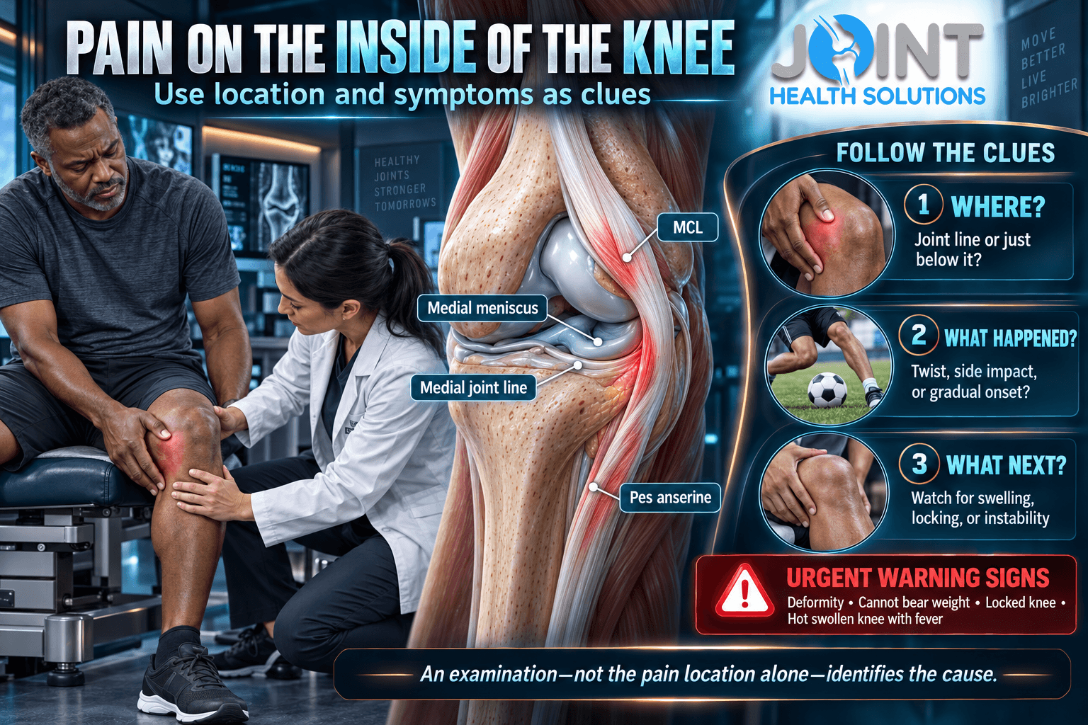
Pain on the inside of the knee—also called medial knee pain—can come from the medial collateral ligament, medial meniscus, inner portion of the knee joint, pes anserine tendons and bursa, nearby bone, or pain referred from another area. The exact spot, how symptoms began, and which movements reproduce them provide useful clues, but location alone cannot establish a diagnosis.
This guide explains why the inner knee may hurt during walking, running, bending, stairs, twisting, or rest; how common symptom patterns differ; which warning signs require faster care; and how clinicians match testing and treatment to the most likely pain source.
Quick Answer: What Causes Pain on the Inside of the Knee?
Common possibilities include a medial collateral ligament (MCL) sprain after a sideways force, a medial meniscus injury after twisting or gradual degeneration, medial-compartment osteoarthritis, and irritation of the pes anserine tendons or bursa a few inches below the joint line. Other causes include medial plica irritation, patellofemoral pain that feels more medial, bone stress injury, fracture, inflammatory or crystal arthritis, infection, and referred pain.
Seek urgent assessment after a significant injury if the knee is deformed, you cannot bear weight, you cannot fully straighten it, or swelling develops rapidly. A hot, markedly swollen knee with fever or feeling ill also needs urgent care. New one-sided leg swelling with chest pain, trouble breathing, fainting, or coughing blood is an emergency—call 911.
What Structures Are on the Inside of the Knee?
The word inside usually means the side of the knee closest to the other leg. Clinicians call this the medial side. Several structures occupy a relatively small area, so two people pointing to “the same spot” may have different pain sources.
The medial collateral ligament runs along the inner side of the knee and helps resist excessive side-to-side movement. The medial meniscus is a C-shaped pad of fibrocartilage between the femur and tibia. It helps distribute load and contributes to stability. The medial compartment is the inner weight-bearing portion of the joint, where articular cartilage covers the bone surfaces.
Below the joint line, tendons from the sartorius, gracilis, and semitendinosus muscles attach to the upper tibia in a region called the pes anserinus. A small bursa reduces friction there. The inner edge of the kneecap, medial plica, hamstring tendons, bone, nerves, and tissues from the hip or lower back can also contribute to symptoms.
Because these structures overlap, self-diagnosis based on one tender point can be misleading. Timing, trauma, swelling, mechanical symptoms, stability, range of motion, and functional loss all change the interpretation.
Inner-Knee Pain Location Clues
Use location as an orientation tool, not as proof of a specific diagnosis. Pain can spread, multiple structures can be involved, and tenderness may occur in more than one condition.
| Pain location | Possible sources | Clues that may matter |
|---|---|---|
| Directly along the inner joint line | Medial meniscus, medial-compartment arthritis, MCL | Twisting, catching, swelling, trauma, stiffness, or weight-bearing pain |
| Along the inner ligament | MCL sprain or tear | Sideways force, tenderness, swelling, or instability after injury |
| Several inches below the joint line | Pes anserine tendon or bursa irritation | Tenderness below the joint, stairs, running, or rising from a chair |
| Inner edge of the kneecap | Medial plica or patellofemoral pain | Pain with repeated bending, stairs, squats, or prolonged sitting |
| Focal pain over bone | Contusion, stress injury, or fracture | Impact, increased training load, night or rest pain, or difficulty bearing weight |
| Diffuse pain extending beyond the knee | Hip, spine, nerve, vascular, or inflammatory source | Back or hip symptoms, numbness, weakness, whole-leg swelling, or systemic illness |
Common Causes of Pain on the Inside of the Knee
No list can diagnose an individual knee. These causes are organized by clinical pattern so you can describe your symptoms more clearly and understand why an examination may be needed.
Medial collateral ligament injury
The MCL can be stretched or torn when a force pushes the knee inward, when the body changes direction abruptly, or during a collision. Symptoms may include tenderness along the inner ligament, localized swelling, stiffness, a pop, or the feeling that the knee is unstable. A mild sprain can remain stable; a complete tear or combined injury may cause substantial looseness.
MCL pain is not confirmed by pressing on the inside of the knee. Meniscus, bone, and joint injuries can accompany the same event. A clinician may compare both knees, assess stability, and decide whether X-rays or MRI would change management. Significant trauma, deformity, inability to bear weight, or marked instability deserves timely assessment.
Medial meniscus injury
The medial meniscus may tear during a pivot, deep bend, awkward rise, or other loaded rotation. Meniscal tissue also changes with age, so a degenerative tear can develop without one dramatic injury. Symptoms may include joint-line pain, delayed swelling, stiffness, catching, reduced motion, or true locking. Many people with a tear can still walk, and an MRI finding does not prove that the tear is the main pain source.
This article targets medial pain location rather than competing with the dedicated guides to meniscus tear treatment and degenerative meniscus tears. Those pages address diagnosis-specific management in greater detail.
Medial-compartment knee osteoarthritis
Osteoarthritis can affect one knee compartment more than another. When the medial compartment is involved, pain may be most noticeable along the inner joint line during standing and walking. Stiffness after rest, reduced motion, intermittent swelling, and changes in walking tolerance may occur. Symptoms and function do not always match the degree of change visible on an X-ray.
Do not assume every new episode is “just arthritis.” Sudden pain after trauma, rapid swelling, true locking, fever, a hot joint, focal bone pain, or new neurologic symptoms require a broader evaluation. For the larger arthritis topic, see the guide to knee osteoarthritis pain.
Pes anserine tendon or bursa irritation
Pes anserine symptoms are generally felt below the inner joint line on the upper tibia. The area may be tender, and pain can worsen with stairs, running, repeated knee bending, or rising from a chair. It may coexist with osteoarthritis or follow an increase in activity. Because the area is below the joint rather than directly within it, careful palpation and movement testing help distinguish it from meniscus or MCL pain.
A label of “bursitis” should not be assumed from soreness alone. Tendon irritation, stress injury, referred pain, and other conditions can create a similar pattern. Persistent symptoms or a sudden loss of function warrant assessment before forceful stretching, aggressive massage, or a generic exercise program.
Medial plica and patellofemoral pain
A fold of joint lining called the medial plica can become irritated and cause pain near the inner edge of the kneecap, sometimes with a snapping or catching sensation during repeated bending. Patellofemoral pain may also be felt around or slightly inside the kneecap, especially during squats, stairs, running, or prolonged sitting.
Clicking alone does not diagnose a plica, meniscus tear, or cartilage injury. The separate article on knee clicking and popping explains why noise must be interpreted alongside pain, swelling, instability, locking, and function.
Bone injury, infection, inflammatory disease, or referred pain
A direct impact can bruise bone or cause a fracture. Repetitive loading can contribute to bone stress injury, particularly when training volume changes faster than the body can adapt. Focal bone tenderness, pain that progresses into ordinary walking or rest, and pain that causes limping deserve prompt evaluation.
A hot, swollen, severely painful joint—especially with fever or feeling ill—raises concern for infection and requires urgent medical assessment. Crystal arthritis and inflammatory conditions can produce a similar acute swollen-joint picture and also require diagnosis rather than home treatment. Hip disease, lower-back conditions, or nerve irritation may refer symptoms toward the knee. New whole-leg swelling, warmth, or discoloration can indicate a vascular problem rather than a knee-structure injury.
What Different Activity Patterns May Suggest
The provoking movement is a clue, not a diagnosis. Use the pattern to guide what you report during an evaluation.
| Pattern | Possibilities to consider | Important accompanying clues |
|---|---|---|
| Pain after a blow to the outside of the knee | MCL sprain, bone injury, combined ligament injury | Swelling, instability, pop, inability to continue activity |
| Pain after twisting or pivoting | Meniscus or ligament injury | Delayed swelling, catching, true locking, giving way |
| Gradual pain with walking or standing | Osteoarthritis, tendon or bursa irritation, bone stress | Stiffness, swelling, focal bone tenderness, changing tolerance |
| Pain with stairs or rising from a chair | Pes anserine, patellofemoral, arthritis, tendon source | Exact location above, at, or below the joint line |
| Pain with deep bending | Meniscus, arthritis, plica, patellofemoral source | Joint-line tenderness, swelling, catching, kneecap-centered pain |
| Pain at rest or at night | Inflammation, advanced joint disease, bone or systemic source | Fever, unexplained swelling, progressive pain, illness, trauma history |
Pain patterns frequently overlap. If the main problem is at night rather than specifically medial, the existing guide to knee pain at night addresses that separate intent. If instability or collapse is the dominant symptom, see the article on knee buckling and giving way.
When Is Inner-Knee Pain Urgent?
Seek emergency or urgent assessment for:
- A visibly deformed knee or kneecap after trauma
- Inability to bear weight or take several steps after a significant injury
- Rapid, large swelling after a pop, pivot, fall, or collision
- A knee that is mechanically stuck and cannot fully bend or straighten
- A cold, pale, blue, numb, or weak foot after an injury
- A hot, markedly swollen or discolored knee with fever, chills, or feeling ill
- New one-sided leg swelling, warmth, or discoloration—especially with recent surgery, immobility, travel, prior clot, cancer, pregnancy, or estrogen exposure
- Chest pain, difficulty breathing, coughing blood, lightheadedness, or fainting with leg symptoms—call 911
Arrange prompt evaluation for persistent swelling, repeated giving way, progressive weakness, focal bone pain, worsening night or rest pain, a suspected MCL or meniscus injury, or symptoms that continue to limit walking and daily activity despite reasonable load reduction.
What Can You Do Before an Appointment?
If there was no major trauma and no warning sign, temporarily reduce the movement that predictably aggravates the knee. This may mean shortening a run, avoiding deep loaded bends, limiting repeated stairs, or substituting a lower-impact activity. Complete bed rest is usually unnecessary for a mild, stable problem, but continuing through a limp or escalating pain is not a useful test of toughness.
A wrapped cold pack may help short-term pain or swelling; protect the skin and use brief intervals. Compression and elevation may be comfortable when swelling is present, provided the wrap is not tight enough to cause numbness, color change, or increased pain. A cane or crutch can reduce load temporarily, but the correct side and height matter.
Medication safety depends on health history, kidney and liver function, gastrointestinal risk, cardiovascular conditions, pregnancy, allergies, anticoagulants, and drug interactions. Ask a qualified clinician or pharmacist before using a new over-the-counter pain reliever. Do not use someone else’s prescription medication.
Track the exact pain location, onset, injury mechanism, swelling timeline, clicking or locking, instability, range of motion, fever or illness, numbness, and which activities change symptoms. This information is more useful than trying to force the knee into a painful self-test.
How Clinicians Evaluate Pain on the Inside of the Knee
Evaluation starts with the story. A direct blow, pivot, gradual activity increase, morning stiffness, fever, previous injury, surgery, clot risk, or pain from the hip or back leads the examination in different directions. The clinician may observe walking, alignment, swelling, skin color, and muscle control, then assess motion, focal tenderness, stability, strength, the kneecap, and the hip or neurologic system when appropriate.
No single office maneuver is perfect. A painful test can be positive for more than one reason, and guarding may make the knee appear less stable or more restricted. Findings should be interpreted together rather than using one press, twist, or click as proof.
When X-rays, MRI, ultrasound, or laboratory tests may help
X-rays can evaluate fracture, arthritis, alignment, and other bone findings, but they do not directly show a meniscus tear or most ligament injuries. MRI may be useful when a significant meniscus, ligament, cartilage, tendon, occult fracture, or bone stress injury is suspected and the result would change management. Ultrasound can assess selected superficial tendons, bursae, fluid collections, or guide certain procedures, but it is not a universal replacement for MRI.
Blood tests, joint-fluid analysis, vascular ultrasound, or emergency imaging may be needed when infection, crystal arthritis, systemic inflammation, or a blood clot is suspected. Advanced imaging is not automatically required for every episode of medial knee pain. The purpose of a test is to answer a clinical question, not simply to produce a picture.
The guide to a first non-surgical joint evaluation explains what information and records may be useful to bring.
How Treatment Changes With the Diagnosis
There is no single best treatment for every person with pain on the inside of the knee. Care depends on the diagnosis, injury severity, stability, swelling, function, goals, medical history, and response to initial management.
| Condition or pattern | Care may include | Important limitation |
|---|---|---|
| Stable low-grade MCL sprain | Protected activity, selected bracing, progressive motion and strengthening | Combined injury or marked instability changes the plan |
| Meniscus-related symptoms | Load modification, rehabilitation, symptom management, selected surgical referral | True locking or a repairable acute tear may need earlier specialist input |
| Medial-compartment osteoarthritis | Education, exercise, weight-management support when relevant, medication review, selected brace or procedure | No nonsurgical option guarantees permanent relief or cartilage regrowth |
| Pes anserine or tendon pattern | Activity adjustment, progressive strengthening, mobility work when appropriate | Confirm the location; joint-line or bone pain may need a different approach |
| Infection, fracture, clot, or vascular concern | Urgent diagnostic and condition-specific medical care | Do not delay care for routine exercise, massage, or an outpatient injection |
Physical therapy and progressive loading
Physical therapy may address motion, quadriceps and hip strength, balance, gait, work or sport demands, and a graded return to activity. The program must match the diagnosis. Exercises that are reasonable for gradual osteoarthritis or tendon symptoms may be inappropriate immediately after a fracture, unstable ligament injury, locked knee, or suspected infection.
Braces, medication, and injections
A hinged brace may protect selected MCL injuries, while an unloading brace may be considered for some people with compartment-specific osteoarthritis. These devices are not interchangeable. Fit, alignment, stability, skin tolerance, and the intended activity matter.
Medication may be considered after contraindications and interactions are reviewed. An injection may be discussed for selected arthritis or inflammatory diagnoses, but pain location alone is not an indication. An injection cannot repair an unstable ligament, mechanically unlock a displaced tear, heal a fracture, or treat a joint infection. Expected benefit, alternatives, evidence, and risks should be discussed for the specific diagnosis. Insurance coverage varies by plan, medical necessity, authorization requirements, and service.
When referral or surgery may be appropriate
Referral may be needed for fracture, persistent mechanical locking, marked instability, a repairable acute meniscus tear, combined ligament injury, progressive neurologic or vascular findings, or symptoms that remain substantially limiting despite appropriate nonsurgical care. A referral does not automatically mean surgery; it helps match the condition to the right level of evaluation.
When Can You Return to Running, Work, or Sport?
A calendar alone cannot determine readiness. The diagnosis, tissue involved, severity, stability, motion, swelling, strength, balance, and task demands all matter. A person returning to desk work has different requirements from someone climbing ladders, kneeling, cutting on a field, or carrying heavy materials.
Progress is generally safer when ordinary walking is comfortable, swelling is controlled, motion is adequate for the task, and strength and control are improving without a delayed flare. Return gradually rather than testing the knee with one maximal session. Pain that becomes sharper, changes gait, produces swelling, or persists into rest indicates that the current load may be too high or the diagnosis needs reassessment.
Evaluation for Medial Knee Pain in Charlotte, NC
Joint Health Solutions evaluates knee pain, swelling, stiffness, instability, injury-related symptoms, and activity limitations at 9350 Benfield Rd, #109, Charlotte, NC 28269. The goal of the initial evaluation is to identify the most plausible pain source and determine whether education, activity modification, physical therapy, imaging, bracing, a selected procedure, or referral is appropriate.
An appointment does not guarantee that an MRI, brace, injection, or other procedure will be recommended. Symptoms suggesting fracture, a locked knee, major ligament injury, infection, blood clot, or circulation problem may require urgent hospital-based care or specialty referral beyond a routine outpatient visit. Call 704-833-3566 for routine scheduling questions; call 911 for a medical emergency.
Frequently Asked Questions About Pain on the Inside of the Knee
What is the most common cause of pain on the inside of the knee?
There is no single most common cause for every person. MCL injuries are more likely after a sideways force, meniscus symptoms may follow twisting or develop gradually, osteoarthritis often produces activity-related pain and stiffness, and pes anserine symptoms are usually felt below the joint line. Age, trauma, swelling, exact location, mechanical symptoms, and examination findings change the probabilities.
How can I tell an MCL injury from a medial meniscus tear?
MCL pain often follows a force that pushes the knee inward and may cause tenderness along the inner ligament, swelling, or side-to-side instability. Meniscus pain is often closer to the joint line and may involve twisting, delayed swelling, catching, locking, or reduced motion. The patterns overlap, combined injuries occur, and an examination is more reliable than self-testing.
Why does the inside of my knee hurt when I run?
Running-related medial pain may involve the pes anserine tendons or bursa, meniscus, medial-compartment joint, MCL, or a bone stress injury. Training volume, hills, speed, surface, recovery, footwear, and previous injury may affect symptoms. Stop running and seek assessment if pain causes limping, becomes focal over bone, progresses into rest, or worsens despite reducing load.
Why does my inner knee hurt when bending or squatting?
Deep bending increases load across the knee and can provoke meniscus, arthritis, plica, patellofemoral, or tendon-related symptoms. Exact location and associated swelling, catching, locking, instability, or trauma help refine the possibilities. A squat response alone cannot identify damaged cartilage or prove a meniscus tear.
Can osteoarthritis cause pain only on the inside of the knee?
Yes. Osteoarthritis may affect the medial compartment more than the other knee compartments, producing inner joint-line pain with standing or walking. Stiffness, intermittent swelling, reduced motion, and declining walking tolerance may occur. Imaging findings should be interpreted with symptoms and function because X-ray severity and pain intensity do not always match.
Where is pes anserine pain usually located?
Pes anserine pain is typically felt on the upper inner shin several inches below the knee joint line. The area may be tender and symptoms may increase with stairs, running, repeated bending, or rising from a chair. Joint-line pain, focal bone pain, swelling, or instability may point toward another or additional source.
Do I need an MRI for medial knee pain?
Not routinely. History and examination come first, and X-rays may be appropriate for trauma, arthritis, alignment, or bone concerns. MRI can help when a significant meniscus, ligament, cartilage, tendon, occult fracture, or bone stress injury is suspected and the result would change treatment. Persistent pain by itself does not make MRI automatically necessary.
Will a knee brace help pain on the inside of the knee?
It depends on the diagnosis. A hinged brace may protect a selected MCL injury, while an unloading brace has a different purpose in compartment-specific osteoarthritis. A poorly chosen or poorly fitted brace may not help and can irritate the skin or alter movement. Bracing should support a defined goal rather than substitute for evaluation.
Can an injection treat pain on the inside of the knee?
Only selected diagnoses may justify an injection, and expected benefits and risks vary. An injection is not a default treatment for medial knee pain and cannot repair an unstable ligament, mechanically unlock a displaced meniscus tear, heal a fracture, or treat infection. A diagnosis-first evaluation should determine whether a procedure belongs in the plan.
When is pain on the inside of the knee an emergency?
Emergency or urgent assessment is appropriate after major trauma with deformity, inability to bear weight, rapid swelling, a mechanically locked knee, or a cold, pale, numb, or weak foot. A hot swollen knee with fever needs urgent evaluation. Call 911 for leg symptoms accompanied by chest pain, breathing difficulty, coughing blood, lightheadedness, or fainting.
Key Takeaway
Pain on the inside of the knee is best understood by combining exact location, injury mechanism, provoking load, swelling, mechanical symptoms, stability, motion, and function. MCL, meniscus, osteoarthritis, pes anserine, plica, bone, infection, vascular, and referred-pain patterns require different responses. Red flags determine urgency; a diagnosis-first evaluation determines treatment.
Sources
- American Academy of Orthopaedic Surgeons: Collateral Ligament Injuries
- American Academy of Orthopaedic Surgeons: Meniscus Tears
- American Academy of Orthopaedic Surgeons: Arthritis of the Knee
- American Academy of Orthopaedic Surgeons: Pes Anserine Knee Tendon Bursitis
- American College of Radiology: Appropriateness Criteria for Chronic Knee Pain
- NHS: Septic Arthritis
- Centers for Disease Control and Prevention: Venous Thromboembolism